
ACOUSTIC SPATIAL ABERRATION CORRECTION
Spatial Distortion and 3D Volumetric Ultrasound Imaging
Wayne Moore, B.Sc., MBA, FASE
April 11, 2017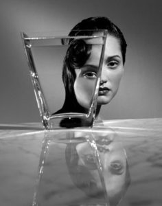
In spite of all the amazing new image processing algorithms and mind-numbingly fast electronics there still remains a significant challenge in clearing up one form of spatial image distortion that persists in current ultrasound images – specifically, the correct positions of the imaged objects are not corrected or maintained. Current spatial compounding techniques work to make point-like targets look point-like. In essence they operate to de-blur the image to some degree. They do not, however, correct for any errors in the positions of those points within the image field.
Diagnostic ultrasound imaging in the body is akin to looking through a shower glass. The image of the person on the other side is both blurred and distorted. The blurring is caused by fairly rapid variations in the glass thickness whereas the distortion comes from relatively slow variations. Current aberration correction techniques tend to remove the effects of the rapid variations, not the slow ones. What results is a fairly well resolved but distorted image much the same as is shown in the photo above, and what one sees through curves and bends in an automobile windshield.
Spatial distortions like the one shown above do not present themselves when one is imaging with a tissue mimicking phantom, as the material used is homogeneous from the top of the phantom to the bottom. Yet when imaging in the human body the “windshield” distortion can sometimes be great enough to cause multiple erroneous features. For example, sonographers and clinicians generally no longer image the womb with the transducer placed along the mother’s midline. The distortion caused by the abdominal muscles in this region are known to give rise to a false impression of twins. Current aberration correction methods do not change this effect and can, potentially, make it worse.
2D matrix array transducer technology has allowed for the capture, processing, and display of 3D volumetric ultrasound images in a display format as shown below. This further complicates the recognition of the effect of slow variation distortions in the resultant 3D image. Volumetric ultrasound images are being used more-and-more in the 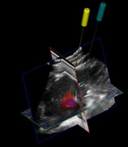 area of image guided intervention, see 3D image below. Exact placement during these procedures is obviously paramount in the successful outcome of the procedure; so, any distortion that could lead to misplacement could potentially be a significant risk to the patient. Additionally, other uses of ultrasound volumetric images are being researched on a daily basis; for example, contrast media imaging combined with chemo-therapeutic treatment, imaging with bio-markers, pre-surgical planning, and 3D printing of the organs, and so on.
Although we have come a long way with both transducer and system technology over the last twenty years we still have some significant design challenges left to create an ultrasound image on par with the resolution of MR and CT scans – but we’ll get there.
About the Author, G. Wayne Moore:
A 30-year veteran of the diagnostic ultrasound market Wayne has held senior level positions with several major medical equipment manufacturers, including Honeywell Medical Systems and Siemens Medical Solutions. Wayne has been directly involved in the development and commercialization of more than 15 technologically intensive ultrasound systems. He is widely published in diagnostic ultrasound literature, a sought after speaker at medical imaging conferences, has served as an expert witness in multiple ultrasound litigations, and holds more than 16 United States ultrasound related patents. Wayne obtained his MBA from the University of Denver – Daniels College of Business.
He was elected as a Fellow of the American Society of Echocardiography (FASE) in 2009.